The eye is not a camera that hands pictures to the brain. Close to forty percent of the brain's nerve fibres are tied to vision: the pathway runs from the retina, through the optic nerve and the thalamus, into the visual cortex — and from there out into the networks for movement, attention, memory and emotion. A shape held steadily at the centre of gaze, expanding and contracting on a slow, even rhythm, is therefore not something merely looked at. It is a way of entering the nervous system at the point where every other system is reachable.
The pathway
Signal pathwayAnatomy
How the signal travels
01
Retina
Cone and ganglion cells translate contrast and edge into spikes. High-contrast geometry produces the cleanest, strongest signal the eye can send — it is the raw material of everything downstream.
02
Optic nerve
Roughly a million axons per eye carry that signal toward the brain. At the optic chiasm the fibres cross, so each shape reaches both hemispheres — which is why a single centred target can engage damaged and intact tissue at once.
03
Thalamus
The lateral geniculate nucleus is the relay and the gatekeeper. Arousal and attention are first folded into the signal here, before anything reaches conscious seeing.
04
Visual cortex
Edges, motion, form and depth are assembled across the cortical stages. A repeating shape drives the same columns in the same order on every cycle, which is what makes the input learnable rather than merely seen.
05
Networks
From cortex the signal branches into the dorsal stream (where it is and how to move toward it), the ventral stream (what it is) and the limbic system (whether it matters). Vision is the shortest route into all three.
06
Plasticity
Repetition is what rewrites the map. Pathways that fire together strengthen, myelin thickens along the ones used most, and networks that had fallen out of step retime to each other. That is the physical basis of rehabilitation.
From the eye to the whole body
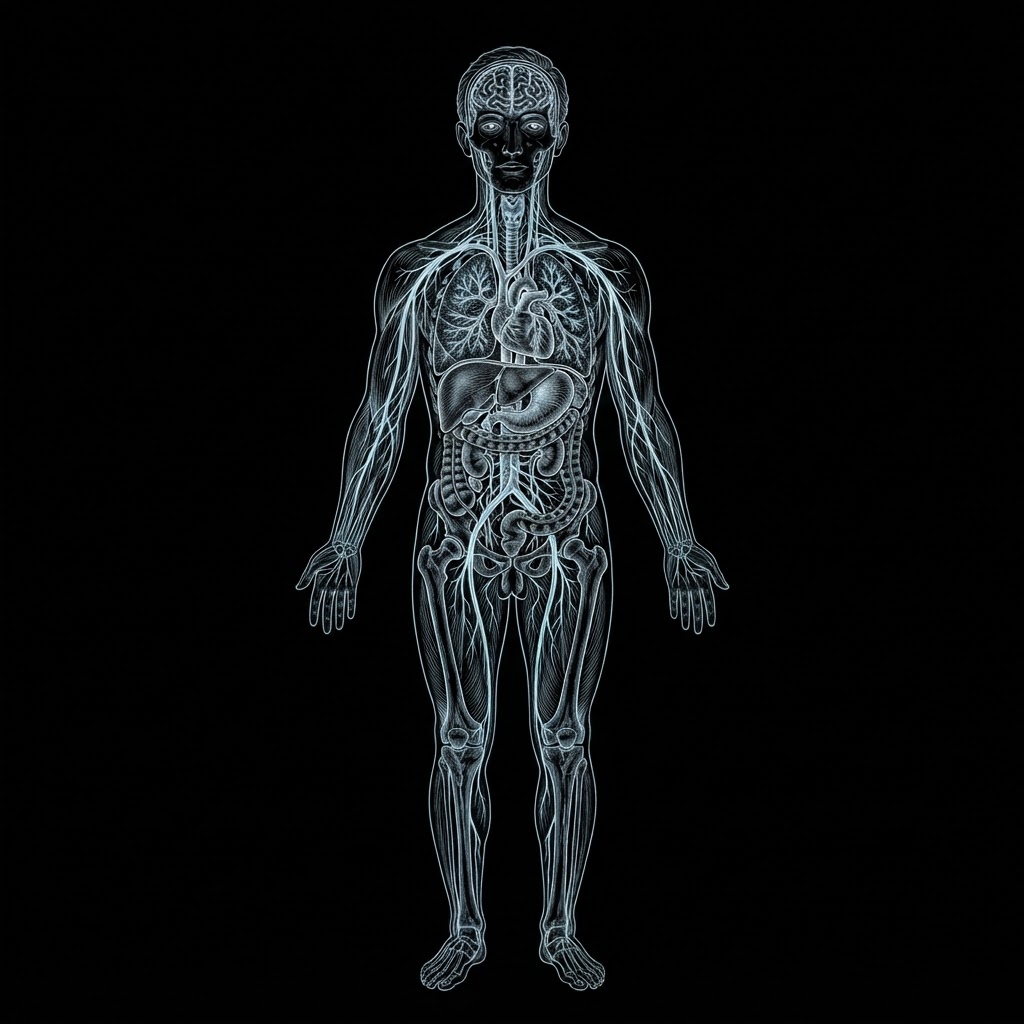
The visual system does not stop at the cortex. Its output descends through the brainstem and spinal cord and reaches the heart, the lungs, the gut, the muscles and the skin — which is why an exercise that only asks you to look at a shape can be felt in the chest, the stomach and the legs. The map below follows that route.
I · The eye — cross-section
Light enters from the left, is bent by the cornea and focused by the lens, and leaves the retina as a spike train along the optic nerve.
1Retina
2Optic chiasm
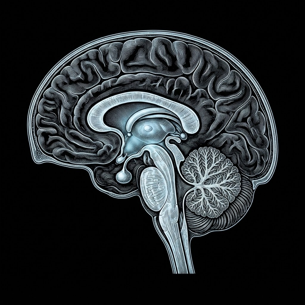
II · The brain — midline section
Every fibre from the eye relays in the thalamus before reaching the cortex — and branches from that same relay descend into the brainstem, the limbic system and the body clock.
3Thalamus (LGN)
4V1 cortex
5Brainstem & vagus
6Cerebellum & balance
7Limbic system
8Hypothalamus & clock
III · The body — where the signal lands
From the brainstem the signal descends the cord and branches into the organs, the skin and the muscles — lungs and heart, liver, stomach and gut, arms and legs.
9Gut–brain axis
10Motor tracts
11Sensory cortex
12Muscles & gait
Anatomical plates — eye, brain, bodyNumbers mark the routes below
1RetinaRods and cones turn the shape into spike trains — the only point where light becomes nerve signal.
2Optic chiasmFibres from the inner half of each retina cross, so each hemisphere receives the opposite half of the visual field.
3Thalamus (LGN)The relay gates how much passes: attention and arousal decide what is allowed through to the cortex.
4V1 cortexEdges, contrast and motion are extracted; the shape becomes a recognised object rather than a patch of light.
5Brainstem & vagusParasympathetic outflow to heart and lungs — a slow, steady gaze lowers sympathetic drive and resting pulse.
6Cerebellum & balanceGaze stabilisation, head position and posture: steady fixation steadies the trunk and reduces dizziness.
7Limbic systemAmygdala and hippocampus tag what is seen with threat or safety. Predictable input lowers alarm and helps memory settle.
8Hypothalamus & clockLight timing sets the body clock; practice at a regular hour anchors sleep–wake rhythm and hormonal release.
9Gut–brain axisVagal tone reaches the enteric nerves, so digestion and appetite follow how settled the nervous system is.
10Motor tractsDescending cortical and tectospinal output sets muscle tone, reaching, hand use and stepping.
11Sensory cortexAttention to a visual target sharpens touch and proprioception — the body map is refined through the eyes.
12Muscles & gaitRepeated gaze-guided movement reprograms walking patterns, endurance and the confidence to move through space.
What it does, condition by condition
The same mechanism is applied differently by each injury. What changes is how much signal the damaged tissue can accept, how stable the input must be, and what has to be relearned around the lesion.
Stroke
What is disrupted
A stroke cuts blood flow to part of the cortex, and with it the fast, synchronised signalling between the eye and the visual, motor and attention networks. Fields of vision can drop, gaze drifts, the body's map of space becomes unreliable, and the simplest targeting task costs enormous effort.
How the shapes engage it
A shape that expands and contracts on an even rhythm gives the retina an unchanging, predictable signal — one a damaged pathway can still lock onto. Retinal ganglion cells and the first cortical stage respond most strongly to contrast and edges, so a high-contrast geometric silhouette recruits the surviving tissue immediately around the lesion instead of asking the brain to search a scene.
How healing is supported
Repetition of the same stimulus is what drives remapping: neighbouring neurons widen their response fields into the territory of the lost tissue, the intact eye-thalamus route re-engages the damaged hemisphere, and the attention networks that steer gaze are retrained to hold a single target. Practised over weeks, this is the mechanism behind visual field recovery, steadier fixation and smoother eye-hand timing.
Seizure
What is disrupted
A seizure is a burst of hypersynchronous firing — a failure of the brain's inhibitory control. Between events cortical excitability often stays elevated, and chaotic or flickering visual input can push a vulnerable network past its threshold.
How the shapes engage it
The exercises use slow, smooth, low-frequency change: no strobing, no rapid contrast reversal, no peripheral flashing. The drive stays inside the range that steadies cortex rather than provokes it, and attention rests on one point of focus instead of sweeping the field — which removes the saccadic overload that precedes many sensory-triggered events.
How healing is supported
Sustained, rhythmic, wholly predictable input reinforces inhibitory tone and the brain's own filtering rhythms, easing the cortex toward the calmer, slower state of relaxed wakefulness. Holding attention on a stable stimulus while arousal stays low is a self-regulation skill: it is practised, not merely watched, and it generalises to the moments before an event.
Brain Injury
What is disrupted
Diffuse injury shears the long white-matter tracts that carry signals between the eye, the thalamus and distant cortical regions. Vision can test as normal while integration, timing and sustained attention are not — the result is visual overwhelm, slowed processing and fatigue after a few minutes of demand.
How the shapes engage it
One moving shape keeps demand low and the target unambiguous: nothing to read, nothing to search, no competing detail. Because the stimulus is predictable, the brain is not forced to re-plan a response every frame; the capacity it saves is spent on transmission, which is precisely what was damaged.
How healing is supported
Short, repeated sessions exercise the same pathway again and again, and repetition is what consolidates white-matter connections and retimes regions that have fallen out of step. Tolerance is the visible measure of recovery: a pattern that fatigued the system in two minutes can, with graded practice, be sustained for ten.
Trauma
What is disrupted
Psychological trauma keeps the threat-detection loop — amygdala, insula, brainstem arousal — running hot. The visual system stays tuned to peripheral motion and sudden change, gaze becomes vigilant and scattered, and the body holds a defensive set that is exhausting to maintain.
How the shapes engage it
Slow geometric rhythm gives the eyes something steady and safe to rest on, with the change confined to the centre of gaze rather than the periphery where threat is normally detected. Predictability is the entire point: the shape does what it did last time, which is the opposite of a danger signal.
How healing is supported
Holding focus on a calm repeating pattern while the breath stays even is a bottom-up route into the autonomic system, the same channel used in somatic therapies. Over time the visual system learns that movement does not mean harm, the startle response softens, and the capacity to stay present with the eyes open — rather than looking away — increases.
What it does for overall health
Because vision is wired into so much of the nervous system, the benefits are not confined to the eyes. They accumulate across comfort, physiology, movement and mood — slowly, and in proportion to how consistently the practice is kept.
Visual comfort
Sustained attention on one expanding shape lets the focusing muscles settle into a single distance instead of hunting across a screen. Blink rate recovers, the tear film stabilises, and the burning, aching fatigue of overused eyes — asthenopia — eases.
Attention and mental stamina
The shape is one object, held still, with nothing to read. Each minute of keeping it centred lengthens the interval before the mind drifts, and that endurance transfers to reading, screens, conversation and driving — the everyday tasks that demand a steady gaze.
A calmer physiology
Slow, rhythmic visual change tends to pull breathing with it. As respiration slows, vagal tone rises: resting pulse settles, heart-rate variability improves and blood pressure stabilises. The body shifts out of sympathetic alertness into a recoverable state.
Balance and posture
Vision is one of three inputs — with the inner ear and body sense — that keep us upright. Practising steady fixation and smooth eye movement sharpens gaze stabilisation, so posture holds with less effort, unsteadiness lessens and the risk of falls falls with it.
Sleep and circadian rhythm
Light exposure is the strongest signal the body clock receives. Practising at a regular hour each day anchors the sleep–wake cycle, making it easier to fall asleep and to stay asleep through the night.
Mood and stress resilience
Predictable, non-threatening visual input calms the limbic circuits that generate alarm. Anxiety and irritability settle, and because the exercise is finite and measurable, it gives a sense of control that sustained illness often takes away.
Movement and daily function
Almost every action is guided by the eyes: reaching for a cup, stepping off a kerb, finding a line of text. As gaze control and visual attention improve, these actions become quicker and safer, and dependence on others for ordinary tasks decreases.
Cognitive engagement
Visual attention is coupled to working memory and the executive networks. Keeping a shape in focus is a genuine cognitive task, and doing it regularly keeps those networks exercised — support for cognitive reserve alongside physical and social activity.
A note: these exercises are a complementary practice. They support — and do not replace — the care of a neurologist, ophthalmologist or therapist, and any change in seizures, vision or sensation should be reported to them.